|
Tumors happen in fish just like in other
humans and other animals. Most of the time they are cancer. Some times in
females they will become egg impacted and are mistaken for cancer. In a good
majority of female fish that become egg impacted they eggs are reabsorbed - but
not always.
I thought my female fish below was egg
impacted. Once I did surgery on her I found she was full of cancer inside.
The pictures below are quite graphic and if
you have a weak stomach now might be the time to hit the "back" button.
|
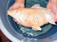
 In the late summer of 2003 I noticed this
gorgeous platinum Ogon getting fat. It
was a little late in the season for her to
be getting eggs. This would be the
first spawn for this 18 inch year old beauty
so I really did not think too much of it to
begin with. The picture was taken with my
underwater video cam.
In the late summer of 2003 I noticed this
gorgeous platinum Ogon getting fat. It
was a little late in the season for her to
be getting eggs. This would be the
first spawn for this 18 inch year old beauty
so I really did not think too much of it to
begin with. The picture was taken with my
underwater video cam.
|

By late December she
continued to grow in size and began to
pinecone. Pine coning is when the
scales stand out on the fish thereby looking
like a pine cone.
I began to suspect she either was egg
impacted or had a tumor. |
I used oil of clove for anesthetic, 5 drops
per gallon of water, and was prepared to
stitch her back up should it turn out she
was just egg impacted.
Note the blood in between the scales and the
general reddish appearance along with the
dissented belly. |
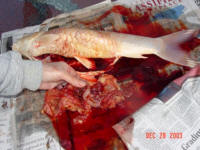
 First cut shows yellowish fluid that had
filled the abdominal cavity. I was
pretty sure there would be no way to save
this beautiful fish now.
First cut shows yellowish fluid that had
filled the abdominal cavity. I was
pretty sure there would be no way to save
this beautiful fish now. |
 A scalpel was used to make the cut from the
pectoral fins back to the anal opening.
A scalpel was used to make the cut from the
pectoral fins back to the anal opening.
|
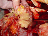
 The yellow mass is the tumor. The yellow mass is the tumor. |
Photos
show that the tumor had encased all the
organs in the abdomen, the gall bladder, the
liver, and the intestinal track I
realized at this point there was no way to
save this fish because removing all the
tumor from these vital organs would be very
tricky indeed, even for them most
experienced surgeons.
|

You can see the swim bladder in the empty
abdominal cavity. Note all the loose
skin from the very large area the tumor
occupied.
 |


